The knee jerk reflex, or patellar reflex, is a cornerstone of any comprehensive neurological examination. It’s more than just a tap on the knee; it’s a window into the integrity of your nervous system, specifically evaluating the L2, L3, and L4 spinal segments. As a classic example of a deep tendon reflex (DTR), its presence, absence, or exaggerated response can provide critical clues to underlying neurological conditions, from peripheral neuropathies to spinal cord lesions.
Let’s explore the fascinating mechanics of this reflex, how to properly assess it, and what various responses can tell us about a patient’s neurological health.
Understanding the Mechanics: The Monosynaptic Stretch Reflex
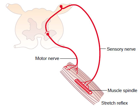
The knee jerk is a prime example of a monosynaptic stretch reflex—one of the simplest and fastest reflex arcs in the human body, involving just two neurons and one synapse.
How it Works:
- The Stimulus: Tapping the patellar tendon just below the kneecap effectively stretches the quadriceps femoris muscle. This sudden stretch is key.
- Sensory Input (Afferent Arc): Specialized sensory receptors called muscle spindles, embedded within the quadriceps, detect this stretch. They send a rapid signal via Ia afferent nerve fibers to the spinal cord at the L2, L3, and L4 levels.
- Direct Connection (Synapse): In the spinal cord, the Ia afferent neuron directly synapses onto an alpha motor neuron in the anterior horn. This direct connection makes it “monosynaptic.”
- Motor Output (Efferent Arc): The excited alpha motor neuron immediately transmits a signal back to the quadriceps muscle.
- The Response: The quadriceps muscle contracts, causing an involuntary extension of the knee – the familiar “jerk.”
Grading the Reflex: The DTR Scale
Deep tendon reflexes are graded on a standardized scale to allow for consistent documentation and interpretation:
- 0: Absent reflex (no response).
- 1+: Hypoactive or diminished reflex (present but less than normal).
- 2+: Brisk, normal reflex (the expected physiological response).
- 3+: Hyperactive reflex without clonus (more brisk than normal).
- 4+: Hyperactive reflex with mild clonus (brief, unsustained rhythmic contractions following the reflex).
- 5+: Hyperactive reflex with sustained clonus (prolonged rhythmic contractions, clearly pathological).

Interpreting Abnormal Reflexes: What Do They Mean?
Variations from the normal 2+ reflex can be highly diagnostic.
A. Hyporeflexia (Absent or Diminished: Grades 0-1+)
A reduced or absent knee jerk suggests an interruption somewhere within the reflex arc, indicating a Lower Motor Neuron (LMN) lesion.
- Common Locations: The problem could lie in the muscle spindle itself, the Ia afferent nerve fibers (sensory nerve), the dorsal root ganglion, or the alpha motor neuron (motor nerve) that innervates the quadriceps.
- Sensory vs. Motor: It’s important to remember that the afferent (sensory) loop is often more critical for reflex function. Unless the muscle is severely paralyzed, reflex loss often points more strongly to a sensory nerve or nerve root lesion rather than a primary motor nerve issue.
- Common Causes: The most frequent culprits include peripheral neuropathies stemming from conditions like diabetes mellitus, chronic alcoholism, specific toxins, radiculopathy (nerve root compression), or severe muscle diseases.
B. Hyperreflexia (Increased: Grades 3+-5+)
An exaggerated knee jerk points to a loss of descending inhibitory control over the reflex arc, typically indicating an Upper Motor Neuron (UMN) lesion.
- Mechanism: Damage to the corticospinal tract or other descending pathways removes the normal “brakes” on the anterior horn cells (alpha motor neurons) in the spinal cord, leading to increased excitability.
- Pathological Significance: While brisk reflexes (3+) can sometimes be non-pathological (e.g., due to anxiety or heightened alertness), the presence of clonus (grades 4+ and 5+) is always pathological in adults. Clonus signifies significant dysfunction of the UMN pathways.
Beyond Hypo/Hyper: Other Distinct Reflex Findings
The knee jerk can also present with unique characteristics in specific conditions:
- Hypothyroidism (‘Hung-Up’ Reflexes): In patients with an underactive thyroid, both the contraction and, notably, the relaxation phases of the deep tendon reflexes are significantly prolonged. This creates a distinctive “hung-up” quality where the leg slowly returns to its resting position.
- Cerebellar Ataxia (‘Pendular’ Reflexes): In conditions affecting the cerebellum, reflexes may appear “pendular.” After the initial reflex, the limb oscillates multiple times, swinging back and forth loosely like a pendulum. This is often associated with reduced muscle tone.
Tips for Eliciting the Knee Jerk Reflex Effectively
- Optimal Patient Positioning: Have the patient sit with their legs dangling freely over the edge of an exam table, or support the knee with your arm if they are lying down.
- Patient Relaxation: Encourage relaxation. Distraction (e.g., asking them to count backward) or the Jendrassik maneuver (having the patient clench their teeth or hook their hands together and pull) can augment reflexes, especially in anxious patients or those with subtle hyporeflexia.
- Proper Hammer Technique: Use a reflex hammer with a brisk, even tap directly on the patellar tendon.
- Observe and Compare: Always test both knees and compare the responses for symmetry. Asymmetry in reflex grading is often a more significant finding than an isolated abnormal grade.
The knee jerk reflex is far more than just a knee-jerk reaction; it’s a sophisticated diagnostic tool, providing immediate insights into the intricate workings of the nervous system. Mastering its assessment is fundamental for any healthcare professional.
What are your go-to tips for eliciting a challenging knee jerk reflex? Share your clinical pearls in the comments below!

Dr. Kunal Varma Bheecarry, MD is a GMC registered substantive Consultant Physician in the UK, specializing in Internal Medicine. With more than 15 years of experience on the clinical frontlines in both General Internal Medicine and Acute Medicine, he is passionate about translating complex medical topics, common problems and clinical guidelines into clear, actionable knowledge for healthcare professionals and patients alike. Every article is written and rigorously reviewed by him and other registered medical professionals to ensure clinical accuracy, guideline compliance, evidence-based reliability, authoritativeness and trustworthiness.
Disclaimer: All content is purely educational and does not constitute personal medical advice. It is not a substitute for professional clinical advice and readers must consult their own primary team. All views and interpretations are mine and not related to the work place.